Friday, April 30, 2021

My XRF Setup - Part 2 / X-Ray Spectrometer

 Amptek (Amptek) is one of the leading companies for Space instrumentation, experimental and research XRF equipment.

They have a fantastic line of products called X-123 Spectrometer - (1) detector element and preamplifier, (2) Digital Pulse Processor and MCA and (3) Power supply.  It is all-in-one device which only requires an external power and connection to a computer. The PC software by Amptek called DppMCA is used to configure, control the X-123 operation and receive & visualize the accumulated spectrum.

Once the acquisition process is started, X-123 doesn't even need the computer connection until it is time to receive, save and display the data, integrated by the internal to X-123 multi-channel analyzer (MCA) located on the DP5 module. 

X-123 Spectrometers are offered with a variety of detectors -  Si-PIN, SDD, Fast SDD or CdTe and can employ different length extenders between the case and the detector element. Fast SDD is their top-of-the-line model, while Si-PIN is more of a general use detector. CdTe detectors are great for the higher energy region - up to 150 keV at the expense of resolution and internal noise.

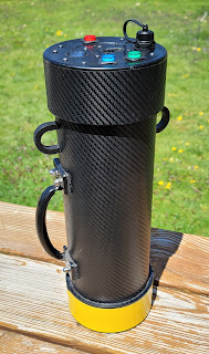
I got my detector from George Dowel (GEO Electronics) as Geo-123. Internally, the unit is identical to the commercial Amptek Si-PIN X-123 unit - George uses the OEM modules and installs them in a custom-machined enclosure. The enclosure is a bit larger than the commercial Amptek version but this is an advantage - the aluminum alloy enclosure actually acts as a giant heatsink for the heat pumped by the TEC module, located inside the detector element and larger surface area results in better heat dissipation.
If there is one thing I wish for, is to have at least 1" or more extension between the detector and the main enclosure - this could help a lot with detector placement in relation to the sample and the exciter.

The "business end" of the unit - the 25 mm2 / 500 μm Si-PIN X-Ray detector element (model FSJ32MD-G3SP) with a thin, very fragile 1 mil Beryllium window.
 
(!) This window must never be touched by hand or come in contact with any object - such thing could turn into a very costly mistake!

Out-of-the-box there is a red polyethylene protective cap installed. There is actually very little reason for the red protective cap to be removed and the detector works with the cap on. I would expect to see some attenuation in the very low end of the range (0 to 2 keV) when the cap is on but even with this cap, Calcium K-lines are actually detectable.
(George supplies a spare modified cap with a built-in thin Kapton window )

Energy resolution is 190 - 225 eV FWHM @ 5.9 keV, peaking time 25.6 μs and Peak-to-Background ratio: 2000/1 (typical).

Plot showing the efficiency as a function of energy for Si-PIN detector. 

The optimal energy range for a SiPIN detector is 1 to 10 keV. 
The range of 10 keV to 25 keV exhibits a drop in efficiency to ~25%. 
Below 1 keV the loses from X-Rays traveling thru the air are significant - only 1cm of air will stop 90% of the X-Rays.
Above 25 keV the detector is still useable up to around 60 keV with a rapidly decreasing efficiency.

The X-123 Spectrometer supports USB 2.0 (mini-USB Connector), RS-232 (2.5mm jack) and Ethernet (RJ45) computer connections. 

USB works just fine and it is very fast so I never had the motivation to try any of the other interfaces. The Ethernet connectivity might require a future software release for full implementation, according to one Amptek document, but the orange data light on the port is a useful indicator - it is lit solid if the data acquisition is stopped and it is blinking when the MCA is running and storing data.
Other connectors on the back are the proprietary jack for the External Power supply and there is also a well documented auxiliary connector for gated counts and other functions.

The "sandwich" of DP5 Digital Pulse Processor (top board) and PC5 power supply module on the bottom. A ribbon cable connects DP5 to the PA230 Pre-amplifier board.

External power is supplied with a very small, proprietary connector (George provides a spare connector in the kit). 
The power adapter is regulated and rated for 5V / 2.5A. 
The current rating is very important - while the unit only needs 500-700 mA during normal operation, there is a short, high-current transient of around 2A during the boot up sequence and any current limiting bellow 2A could damage the internal power supply PC5 module.
All of the power conditioning and the generation of various voltages is done internally by the PC5 power supply module.

The Amptek software - DppMCA is quite good and I really like it! It is available on the AmpTek web site for free. The software is fairly easy to use and provides extensive toolset for data acquisition and analysis. The peak identification feature using energy reference libraries is very useful. The UI is logical and easy to use and ability to customize the color schemes.

There are a few features I wish it had but overall it does its job very well and it is well integrated with the hardware DP5 Pulse Processor.
Speaking of the DP5 module, the built-in hardware MCA in X-123 is quite impressive - 256 to 8192 channels (I normally use it in 4096 channels configuration) and 24 bits per channel (16.7 million counts). Acquisition time is selectable from 10 ms to 466 days. 
The MCA can be set to work in two modes - NORMAL and DELTA. In Delta mode it shows the spectrum, refreshed every second with pulses integrated over the past 1 second.

Combination of coarse and fine amplifier gain yields an overall Gain, continuously adjustable from x0.84 to x127.5 - the amount of preamp Gain determines the spread of the spectrum over a specified number of channels in the MCA.
For example, when using 4096 Channels, a Gain of x18.5 allows coverage of 0 to 62 keV range.

The Si-PIN detector response is quite linear and 2 point calibration is all that is needed for most applications.
I use pure, 99.9% Copper (Cu) foil - the Kα1 line at 8.05 keV and the Am-241 X-rays at 59.54 keV at the high end of the spectrum are sufficient for channel/energy calibration but more intermediate points can easily be added if necessary using different pure metals.

Gadolinium (Gd) is a Rare-Earth Element which is very interesting to XRF with its many peaks and also can be used as a calibration aid since both L and K-lines show up nicely at the low and high energy range of the detector.

XRF of a 99.9% pure 1" disk of Gadolinium (Gd).

Most of the Gd peaks can be easily identified - Kα1, Kα2, Kβ1, Kβ2, Lα1/Lα2, Lβ1, Lβ2 and even Lγ1, Lγ2 and Ll are visible in this plot. Obviously, Lα1 and Lα2 can not be separated - they are only 30 eV apart - way too close for the 190-225 eV resolution of the Si-PIN detector.

The very low count "hash" above the group of Gd L-lines is caused by Np-237 L-lines coming from the native spectrum of the exciter - 25 μCi of Am-241. Am-241 decays to Np-237 and the L-lines of the Neptunium are Rayleigh scattering and somewhat visible in the spectrum. 
The Exciter's X-Ray beam is collimated and reduced down to about 3mm spot for a precise sampling so the overall count rate is low as expected but the spectrum is nice and fairly clean. Longer integration times are to be expected with this type of exciter and I use a different exciter with a broader beam for general purpose.

Thursday, April 29, 2021

My XRF Setup - Part 1 / How it all works?

 What is XRF? 

XRF stands for "X-Ray Fluorescence" and there are two main types: Energy Dispersive XRF and Wavelength Dispersive XRF. 

I'll focus on the Energy Dispersive, Direct Excitation (2D) Method as this is what I use. 

EDXRF is a Non-Destructive method for material analysis, used to determine the Elemental composition of a material or a chemical compound. It is an extremely useful tool to analyze raw materials, minerals, alloys, etc.

The Physics behind XRF is absolutely fascinating and at the same time relatively  simple to understand.

The description of the whole process can be boiled down to this: exposing a test sample to a beam of X-Ray radiation and detecting the energy of the secondary / characteristic X-Rays emitted by the atoms in the sample and then building a histogram of the energy spectrum in order to identify specific secondary X-Ray peaks.

When a material is irradiated by short-wavelength ionizing radiation like X-Rays or low-energy Gamma Rays, the electrons from the innermost electron shells, the ones closest to the nucleus (K, L, M shells) will become excited and are expelled from the atom. This causes a vacancy in that lower electron shell and it is immediately filled with an electron from a higher-energy shell.

For example, if the electron is ejected from the K-shell, this vacancy will be filled by an electron from the L or M shells. When the electron makes the jump from a higher-energy shell to a lower-energy shell in order to fill such vacancy, it must give off the excess energy and does it so by emitting a photon with energy equivalent to the difference. This secondary photon is again, an X-Ray photon but with a very specific energy to the particular element due to the unique binding energy between the nucleus of each element (with its protons) and the surrounding electron shells. 

This, secondary emitted photon is called a Characteristic X-Ray. By detecting the energy of these characteristic X-Rays we can determine which Element from the Periodic Table the examined atom belongs to. 

There is a number of such characteristic X-Rays emitted, based on which shell, the electron comes from to fill the vacancy of the electron expelled by the primary X-Rays - if the vacancy is in the innermost shell (K-shell) and it is filled from the L-shell it is called Kα energy, if it is filled by the M shell is Kβ energy and so on. Vacancies in the L-shell are filled from the M-shell and are called Lα and when filled from N-shell - Lβ

These characteristic X-rays energy are published in lookup XRF tables.

Overlaps between Kα/Kβ and Lα/Lβ energies for some elements exist so identifying an element often relies on identifying multiple energy peaks in the spectrum, coming from different transition lines.

This is a typical XRF histogram as produced by the MCA software. I used a small sample of pure (99.98%) Cobalt metal for this test. The two blue peaks on the very left are the Kα and  lines of Cobalt. 

The other peaks in the spectrum above are just parasitic peaks coming from the X-ray exciter or the environment - for example the tall green peak on the right of the Cobalt peaks is the Bromine 1-line @ 11.92 keV immediately followed by the Br 1-line @ 13.29 keV, all coming from the plastic clamp I used to hold the small Cobalt metal sample in front of the detector - the Bromine was likely used in manufacturing of the dye or filler of the plastic and even though the clamp was just partially exposed to the detector the Bromine (Br) lines were still detected.

What do I need for XRF?

At a very basic level - three things : X-Ray Source, X-Ray detector and a computer.

The Amptek XRF Kit - Mini-X X-Ray tube and the all-in-one X-123 detector.

X-Ray source: obviously the best source is an X-Ray tube in the 40-60 kV range but these require forced cooling, HV power supply, lots of shielding and a collimator. Such setups are large and very expensive. There are small and portable tubes but they usually have an even higher price tag.

Alternatively a radioactive isotope emitting low-energy gammas / X-Rays can be used as an exciter - Cd-109, Fe-55 or Am-241 just to name a few. The intensity is usually lower than the beam from an X-Ray tube, even when mCi amounts of activity are used so counting times are longer, but it is a very portable and uncomplicated method to produce the primary X-Rays. It is important that the energy of the exciting X-Ray beam is higher than the characteristic energy to be detected.

Needless the say, regardless whether the exciter is an X-Ray tube or a Radioactive Isotope, caution must be exercised at all times dealing with ionizing radiation. 

X-Ray Detector: the X-Ray detector must have very high-resolution (typically 122-200 eV or less) as some peaks are really close to each other and high efficiency in the low-end of the X-ray energy spectrum - typically, efficiency is >25% in the 1 to 25 keV range but my detector covers energies all the way up to 60+ keV at a reduced efficiency.

The required resolution and energy range are normally outside of the capabilities of most Gamma Spectroscopy detectors. Even the thin-crystal GS probes designed for the X-ray region will not have the resolution needed - some limited XRF might still be possible though.

 A specialized semiconductor X-Ray detector device is needed - Si-PIN, SDD or CdTe detector.

These detectors are very expensive, complex and very delicate devices employing a thin and very fragile (0.5 mil or less) Beryllium window and are usually evacuated or filled with low-pressure Helium gas. Inside the detector device are housed many components: the Si-PIN or SDD detector semiconductor chip, an input FET transistor for the preamp, a temperature sensor, a built-in multi-stage thermo-electric cooler (TEC) with a delta of ~85°C which reduces thermal noise in the detector chip and a temperature sensor. The heat pumped out from the chip must be constantly dissipated in the environment thru the mounting stud and the component's back surface thermal interface.

The detector is connected to a charge-sensitive pre-amplifier and the output of the pre-amp is fed into a Digital Pulse Processor (Dpp) which does the pulse detection, pulse shaping, ADC and pulse-sorting as it has a built-in Multi-Channel Analyzer (MCA) (8k channels). 

A Power Supply module generates the bias for the detector, the power to the TEC module and controls the temperature of the detector chip, besides powering the pre-amp and Dpp.

Because of the very low Characteristic X-Ray energies of light elements it can be extremely difficult to detect these elements as their secondary X-rays are easily absorbed even by air - lightest elements emit energies <1keV.

 High-Intensity primary x-ray beam, very thin Be-window, Silicon-Drift Diode (SDD) detector, vacuum chambers and even Helium-filled test chambers are often needed for elements lighter than Potassium to be detected.

Typical Si-PIN detectors work well for elements heavier than Scandium (Z>21) but I am actually able to observe even the Calcium lines - not in great detail but visible in the spectrum.

Tuesday, April 20, 2021

Gamma Spectroscopy - Lead Shielding / Castle v2

I decided to rebuild my Lead Castle / Test chamber for Gamma Spectroscopy. 

The old vertical Lead Castle was working great but after awhile it became evident  that the design is not the most convenient one, especially if it is used often - every time I needed to access the test chamber, I had to partially disassemble and remove most of the lead modules (which are also quite heavy) and then put everything back together. 

Horizontal design is a more convenient option and creates a more accessible test chamber on the expense of the desktop footprint.

I redesigned the castle so I can use some components from the old castle - the detector is also in a horizontal position and can stay in place while I am only manipulating the sample and the end-cap shielding.

There are 3 main shielding components - outer sleeve, inner sleeve and end-cap.


For the new design I reused the main shielding sleeve from the old chamber - 6" long Schedule 40 PVC pipe with 4" diameter. There is about 0.5mm of copper foil wound directly around the plastic form and almost 1" of Lead (rolled sheet with 1/8" thickness). 
The whole package is tightly wrapped with duct tape. The overall outer diameter is 6". 
The outer shield doubles the amount of lead around the test chamber and the detector's NaI(Tl) crystal in this configuration.

For the inner shield I used 12" long section of 2" copper pipe and wound 5 turns of 1/8" thickness Lead sheet. Again, everything is tightly wrapped and covered with duct tape. 

The inner shielding is twice as long as the outer sleeve (12" vs 6") and shields the length of the entire detector assembly.

I built a special, custom wooden crate to hold the lead shielding. The base is made of 3/4" pine 6" x 16". The two sides around the large lead sleeve are glued and screwed with 2" 1/2 long decking screws to the base. 
Two threaded rods, inserted in transparent vinyl tubing, together with washers and nuts re-enforce the top part, maintain the spacing of the sides while allowing the outer sleeve to be removed from the top after the inner sleeve is removed thru the front or the back. The front threaded rod (which also has a larger diameter) doubles as the front carry handle.

The rear portion is a boxed off extension and it has 3/4 pine raiser on which the inner sleeve lays on so it aligns with the outer sleeve. A second carry handle is attached to the top of this box.
Assembly takes place in reverse order. Outer sleeve is dropped from the top into place and then the inner sleeve is inserted.

The 6" long second box section sits on top of the base and the inner sleeve rests on it. It is attached to the base from the bottom side with the same long decking screws and wood glue. A 1" "lip" is left on the back to support a lead shield cap. 

This is the end through which the GS detector is inserted and the detector position is adjusted inside the copper tube lining in order to place the front face of the crystal right against the sample.

The front end of the chamber with the inner sleeve nested inside the outer sleeve. The white front face of the detector is visible inside the copper lining. 
The bottom of both compartments is lined with 1/8" rubber lining. The threaded rod on top is a structural components as the whole crate becomes very heavy (~65 Lbs.) and it acts as a handle as well.

After the test sample is placed in the chamber, a cover made of 6"x 6 x 1/4" Lead plate and 6" x 4" x 1/8" copper plate (towards the chamber) is used to close the opening of the chamber and another 1.5" inches of lead bricks are stacked. The front lead cap on the detector side has an opening for the coaxial cable.

The complete lead castle (v2). 4 rubber feet are installed on the bottom of the base.
While this design puts slightly less lead (5-7mm less) around the crystal than the vertical design (with cast inner shielding), it is much more convenient to use and operate.

Two coats of polyurethane varnish as a finishing touch.